
, by Nadia Jaber
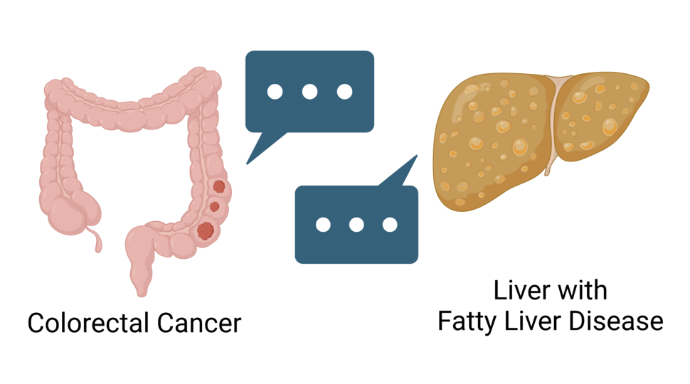
Fatty livers send “message bubbles” to colorectal cancer as it spreads, encouraging the cancer to grow in the liver and preventing the immune system from attacking cancer in the liver.
Credit: Created with BioRender.com
Colorectal cancer often spreads to the liver, and once it does, it becomes incredibly difficult to treat. Cancer is even more likely to set up camp in the liver when patients have fatty liver disease, but scientists don’t know why. Now, a new study has shown that fatty liver disease appears to create the perfect environment for metastatic colorectal cancer to thrive in the liver.
Fatty liver disease happens when excess fat builds up in the liver. Around 25% of US adults have the most common type of fatty liver disease, called nonalcoholic fatty liver disease (NAFLD), and that number is going up. In addition to being linked with colorectal cancer metastasis, NAFLD is also associated with an increased risk of several cancers, including liver cancer.
The NCI-funded study, largely conducted in mice, found that fatty livers sent fat-coated “message bubbles” to colorectal cancer cells. The message bubbles, called extracellular vesicles (EVs), encouraged colorectal cancer to grow in the liver and prevented immune cells from attacking metastatic tumors in the liver. The findings were published May 9 in Cell Metabolism.
“What this study shows is that NAFLD can impact the aggressiveness of metastatic cancer—specifically of colorectal cancer that metastasizes to the liver,” said Joanna Watson, Ph.D., of NCI’s Division of Cell Biology, which partially funded the study.
The findings also raise the possibility that people with colorectal cancer and fatty liver disease might need different treatments than people with colorectal cancer and healthy livers, said the study’s lead scientist, Ekihiro Seki, M.D., Ph.D., of Cedars-Sinai Medical Center in Los Angeles.
Extracellular vesicles from fatty liver
The cells in our bodies can’t whip phones out of their pockets and text each other when they need to communicate. But they can send out EVs, which can travel through the blood to deliver messages to cells in other parts of the body. The messages are typically encoded by proteins or RNA tucked inside the vesicles.
Countless studies have shown that cancer cells rely on EVs to sweet-talk other cells, often as a means of getting their way. Among other things, tumor-derived EVs have been found to tell distant organs—like the liver—to get ready for the cancer’s arrival.
But little attention has been paid to EVs produced by the liver itself and whether they interact with cancer as it spreads, Dr. Seki said. The research team was curious whether EVs from fatty liver disease might help colorectal cancer spread to the liver.
First, the team studied mice with fatty livers. Colorectal cancer cells injected into the spleens spread and formed more tumors in the livers of mice with fatty livers than in those without, they found.
Mice with fatty livers also had more EVs in their blood than their counterparts without fatty livers, the researchers found. Likewise, people with NAFLD had higher levels of EVs in their blood than in people without NAFLD.
EVs from mice with fatty livers and people with NAFLD appeared to tell colorectal cancer cells to grow and spread. For example, when the researchers took those EVs and mixed them with colorectal cancer cells in lab dishes, the cancer cells grew aggressively.
But in mice that were genetically engineered to lack the ability to make EVs in their livers, colorectal cancer cells formed far fewer metastatic liver tumors than in mice with livers that could make EVs.
These findings are novel, Dr. Watson said, because “they focus on liver-produced extracellular vesicles rather than cancer cell–produced extracellular vesicles to create the pro-metastatic niche” (an area in a distant organ that has been prepped to support metastatic cancer growth).
A message for cancer cells and immune cells
The team’s next question was: What’s the message EVs from fatty livers are carrying?
The message, they discovered, came in the form of microRNAs, small pieces of RNA that limit or stop the production of specific proteins in cells. They identified three microRNAs that launched a cascade of actions in colorectal cancer cells that ultimately helped the cells grow and form tumors in the liver. Turning this cascade off in colorectal cancer cells slowed the growth of tumors in mice with fatty livers.
But that’s not all this cascade—controlled by a protein called YAP—appeared to do. It also prompted colorectal cancer cells to alter immune cells in the liver, the researchers reported. More specifically, the cancer cells convinced nearby immune cells not to attack metastatic tumors in the liver.
For example, in mice with fatty livers, colorectal cancer cells released a protein that made immune cells called macrophages migrate into the liver. These macrophages, in turn, sent signals that encouraged cancer cells to grow and cancer-killing T cells to stand down.
Nonalcoholic fatty liver disease and cancer
The next question was: Are EVs from the livers of people with colorectal cancer and NAFLD doing the same thing? To get at the answer, the researchers examined colorectal cancer tumors that had spread to the liver in 16 people with and 18 people without NAFLD.
In people with NAFLD, the YAP signaling cascade was turned on in metastatic liver tumors. There were also more cancer-friendly macrophages and T cells in the metastatic liver tumors of people with NAFLD.
Because NAFLD generally doesn’t cause any symptoms, it “tends to be diagnosed by chance as a consequence of tests for other health problems,” Dr. Watson noted.
Doctors tend to look out for severe forms of fatty liver disease like cirrhosis, Dr. Seki added, and pay less attention to the mildest form, simple steatosis.
“However, I want to tell clinicians, please do not ignore simple steatosis, which can also increase the risk of [certain] cancers and cancer metastasis,” he emphasized.
Not only that, but the study suggests that fatty liver disease might also affect how certain cancer treatments work, Dr. Seki said. Given the immune cell changes they observed, the researchers “strongly expect” that immunotherapies may not work as well in people with fatty liver disease, he explained.
The good news is that fatty liver disease may be improved by losing weight, switching to a low-fat diet, and exercising, he added. And researchers are testing potential drug treatments for NAFLD.
The study also points to the need for a shift in how cancer research is done, said Dr. Watson.
Scientists know “a tremendous amount about the changes that occur within the cancer itself” and the microenvironment right around the tumor, she noted. But scientists also need to think about the “macroenvironment” of the whole body, and how multiple organs and systems are in communication with tumors, she emphasized.


")
