 Shutterstock / David A Litman
Throughout its life cycle, a normal cell will divide around 50 times before it stops, at which point it dies and get replaced by a new cell.
But not all cells die when they stop dividing. Instead, they continue to age without either dividing or dying, meaning they begin to accumulate in tissues.
This is a process called senescence. Senescence can be caused by a variety of stresses on the cell, from DNA damage to exposure to radiation.
Unfortunately, senescent cells don’t accumulate harmlessly. Instead, they take on what’s known as a senescence-associated secretory phenotype (SASP), meaning that they start to secrete substances that are harmful to the cells around them.
As such, senescence is associated with a decline in tissue function as we get older. The more senescent cells we have, the less our tissues can function normally.
Initially, we thought that senescence was our body’s way of suppressing cancer by preventing these damaged cells from dividing and forming a tumour.
However, research has shown that the opposite can also be the case. The senescent cells can promote tumour formation, which makes them a prime target for cancer prevention and interception.
The problem is that studying senescent cells in vivo, meaning in living organisms, has proven very difficult. They can be hard to identify, and they can differ depending on what type of cell they are, where they are in the body, and what stressors have caused them to become senescent.
To change that, researchers led by Dr Scott Haston at the University College London Institute of Child Health and the Early Cancer Institute at the University of Cambridge, with funding from us, have developed a new model to study senescence in.
And what’s more, they’ve found that clearing a certain type of senescent cell could prevent some lung cancers.
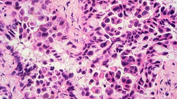
Cherry red senescent cells
From studying senescent cells in vitro, meaning grown in a petri dish or test tube for example, we know that they produce a protein called p16.
p16 is a specific type of protein called a cyclin-dependent kinase inhibitor (CDKi). Cyclin-dependent kinases are a family of proteins in our bodies that are responsible for regulating the cell cycle, that being the process by which our cells grow and divide in a controlled manner.
By inhibiting CDKs, p16 effectively pauses the cell cycle, preventing cells from dividing.
So, to study senescent cells in a living organism, we need to be able to track when p16 is produced. The team did that by modifying the gene that produces p16, called CDKN2A, so that when p16 is produced, another protein called mCherry is also produced.
mCherry is a fluorescent protein that shows up as bright red when exposed to certain wavelengths of light.
And that proved successful.
The team were able to identify mCherry, and therefore p16, in the mice. That gave them a way of accurately pinpointing and tracking senescent cells.
In doing so, they were able to see that the number of senescent cells across tissues was greater in older vs younger mice, consistent with an accumulation of these damaged or dysfunctional cells during the ageing process.
But creating a way to track senescence in vivo was only the first piece of the puzzle.
“We decided to make a mouse model,” says Professor Juan Pedro Martinez-Barbera, professor of developmental biology and cancer and one of the co-lead researchers of this project.
“We made it. And when we had one, then we thought, ‘okay how can we validate it in the context of cancer?’”
An unexpected result
To do that, the researchers decided to use this method of tracking senescence in another group of mice, those with lung cancer caused by mutations in a gene called KRAS.
KRAS is a type of gene called a proto-oncogene. That means that when it’s mutated, it becomes an oncogene, a gene that has the potential to cause cancer. Mutations in KRAS are implicated in several cancers, namely lung, colorectal and pancreatic.
By tracking senescent cells in a mouse model of lung cancer, the researchers hoped that they could pinpoint which tumour cells were senescent so that we know which cells could act as potential drug targets.
But one thing came as a surprise to the researchers. They expected that, although they weren’t dividing, they would find some of these senescent cells inside the tumour, but they didn’t.
“We started with hypotheses that some tumour cells would become senescent, and then we were going to be able to eliminate them to determine their effect in tumour formation,” says Martinez-Barbera.
“That could not have been more wrong, because when we induced the tumours, we saw actually the tumour cells were doing what tumour cells usually do, divide rapidly.”
Instead, they found that senescent cells were in the tumour microenvironment, an interconnected system of cells like blood vessels and immune cells around a tumour that it needs to keep growing.
The microenvironment also contains a lot of immune cells, and it was one specific type of immune cell that was proven to be senescent.
“Surprisingly, we identified these senescent cells as macrophages,” says Dr Daniel Muñoz-Espín, research group leader at the Early Cancer Institute and co-leader of the research team.
“That was the first surprise, and the other one was that when we removed these cells, we found that the tumour burden was strongly reduced in the mice, and the survival increased. Significantly increased.”
Stopping the start
By secreting the harmful substances typical of senescent cells, the macrophages were making the lung tumours worse. And if they were removed, the tumour burden, the number of tumour cells present, fell dramatically.
But these macrophages may be doing more than just helping a tumour once it’s started growing.
When a mutation occurs in the KRAS gene, the cells don’t automatically become cancerous. Instead, they form what are known as pre-cancerous lesions. These lesions have the potential to become cancerous but may not without an external stimulus.
If these macrophages accumulate in these lesions, and begin secreting harmful substances, they may be what’s triggering tumour formation in the first place.
Therefore, if we can identify high-risk patients and test for KRAS mutations, we may be able to stop a lung tumour from forming.
“This can potentially be used as a preventative therapy, because if we eradicate these cells, we can prevent cancer onset, reduce the tumour burden and progression, and increase the survival, at least in our preclinical models,” says Muñoz-Espín.
It might sound like there’s a catch there. Mouse models, though a good way of studying tumour biology, don’t exactly match human cancer biology, so this pattern of senescence may not directly translate into humans.
Luckily, the team have that covered.
By looking at samples from human patients they found that this population of senescent macrophages was present in premalignant lung lesions, which shows that these cells might also be driving lung cancers in humans.
“We’ve shown that there is a population of macrophages in these early lesions in human patients,” says Martinez-Barbera. “And that this population of macrophages was expressing similar markers to what we have seen in mice. So that was very exciting.”
The future of prevention
Although many people may have lesions of KRAS mutated tissue in their lungs, only a minority of them ever develop lung cancer. And predicting who in this population will develop cancer has proven a challenge.
This research shows that if we can identify and remove these senescent macrophages, we may be able to prevent certain lung cancers in the highest-risk population, or to intercept lung cancer at early stages of its development.
“I think this can be potentially an opportunity for these high-risk patients, like those with a long smoking history that’s resulted in multiple precancerous lesions, because they usually they don’t have any access to any available therapy,” Muñoz-Espín explains.
“The only option currently is regular monitoring by CT or by X-ray imaging every six months because chemotherapies, radiotherapy and other cancer targeted therapies usually work in tumours where the cells are actively dividing.
“And it’s not possible to remove these lesions surgically as these patients may accumulate too many.”
So, while this research is still in early stages, it could provide hope for an effective way to not only treat, but to intercept and prevent lung cancer, one of the biggest cancers of unmet need.
Jacob


")
